INFORMATION AVAILABLE IN ENGLISH, GUJARATI AND HINDI
What is a haemangioma?
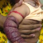
A haemangioma is a collection of small blood vessels that form a lump under the skin. They are sometimes called ‘strawberry marks’ because the surface of a haemangioma looks a bit like the surface of a strawberry.
Haemangiomas can be superficial or deep. Some haemangiomas are a combination of the superficial and deep kinds, with a raised, red area on the surface of the skin, and a bluish swelling of abnormal blood vessels deeper in the skin.
What causes haemangiomas?
There is now some scientific evidence to suggest that nearly three quarters of haemangiomas may be caused by placental tissue. If a tiny piece of tissue from the placenta embeds into the baby’s developing tissue very early in pregnancy, this can start to grow on his or her skin after the baby is born, forming a haemangioma. We do not yet know fully what might cause the placental tissue to embed. Other theories have evolved, but more research is needed to confirm the causes of haemangiomas.
How common are haemangiomas?
About one in every ten babies has a haemangioma. They are more common in girls, in premature babies, low birth weight babies and multiple births, such as twins, triplets and quadruplets. Haemangiomas are not inherited, but because they are quite common, families often report another relative who has had a haemangioma in childhood.
What do haemangiomas look like?
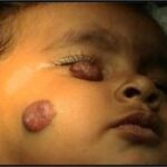
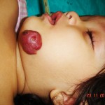
Superficial haemangiomas are usually a raised, bright red area of skin, which feels quite warm because the abnormal blood vessels are close to the surface. A superficial haemangioma may appear initially as a small area of pale skin on which a red spot develops.
Deep haemangiomas may appear bluish in colour because the abnormal blood vessels are deeper in the skin. Sometimes they are not noticeable for the first few weeks, only appearing as a lump as the haemangioma grows; this is particularly true if the surface of the skin is not affected.
Haemangiomas are not usually present at birth but develop a few days or weeks later. They often grow rapidly in the first three months, increasing in size and sometimes in redness. It is unusual for haemangiomas to grow after six to ten months of age, when most haemangiomas tend to have a ‘rest period’ and then begin to shrink.
Where do haemangiomas occur?
Most children only develop one haemangioma, but occasionally, a child has multiple haemangiomas in various parts of the body. This seems to happen most often in twins or other multiple births.
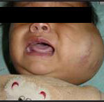
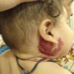
Haemangiomas can appear anywhere on the body. The majority of haemangiomas appear on the head or neck, particularly on the cheek, lips or upper eyelid. Haemangiomas can also appear on the organs inside the body. This is most common when a child has multiple haemangiomas. The organ most commonly affected is the liver, but occasionally the airway, heart and brain may also be involved. Babies with multiple haemangiomas should have an ultrasound scan to confirm this or rule it out.
How are haemangiomas diagnosed?
Superficial haemangiomas are clearly noticeable and quite different to other types of birthmark so no special diagnostic tests are usually needed.
A child with a haemangioma near the eye, or a deep haemangioma, or one affecting the internal organs may require ultrasound and/or MRI scans to confirm the diagnosis, location, and also to check the depth of the affected blood vessels.
Looking after your child’s haemangioma
In most cases, haemangiomas just need looking after carefully.
As the blood vessels in a haemangioma are so near the surface of the skin, they can bleed if they are knocked or scratched. It is important to keep both your nails and your children’s nails cut short and buffed smooth, so that they don’t catch the surface of the haemangioma.
If the haemangioma starts to bleed, apply pressure over it with a clean handkerchief, cloth or tissue for at least five minutes. If blood soaks through the handkerchief, cloth or tissue, put another one on top and keep up the pressure. Do not take it off to have a look as this could start the bleeding again. If the bleeding continues, even after pressing down on the haemangioma for five minutes, go to your nearest doctor or contact us at the numbers provided.
The surface of the haemangioma is delicate and can be very dry, so avoid using bubble baths and rinse any soap or shampoo off carefully and pat the area gently afterwards. A thin layer of coconut oil or jelly (Vaseline etc.) put gently over the top of the haemangioma can stop it drying out.
Haemangiomas need protection from the sun because it can make them swell up for a while and look redder. You can use sun protection cream on all areas of exposed skin or use a hat to protect your child’s face and/or an umbrella when the baby is outdoors.
When might my child’s haemangioma need treatment?
Most haemangiomas do not require any treatment, but there are circumstances when treatment might be needed.
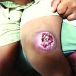
Occasionally, haemangiomas can form an open sore or ulcer, which is painful. Ulcers can become infected, so a visit to your doctor is important, as infected ulcerated areas may need treatment with antibiotics.
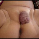
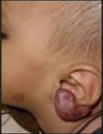
Haemangiomas that are around the mouth, in the nappy area or in natural folds of skin, like the armpit, ear and neck, are most likely to become ulcerated. This is often due to the friction of the two surfaces of skin rubbing together.
A child with a haemangioma around the mouth can suck or chew the lip/ haemangioma, and feeding, either by breast or bottle, can irritate the area further.
The nappy can rub haemangiomas in the nappy area, and contact with stools or urine makes the ulcer more painful. Wherever the haemangioma is located, an ulcer is painful, more likely to bleed, and more likely to become infected. In the long term, an ulcerated area is more likely to leave a scar than a non-ulcerated area of haemangioma.
If your child’s haemangioma develops an ulcer, it will need special attention until it heals. Keep the area clean by washing it twice a day, preferably in a bath or by pouring water over the area, and leave it to dry naturally.
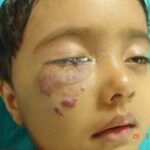
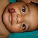
Haemangiomas near the eye
Haemangiomas near the eye can have long-term effects on a child’s vision, so need to be checked by a specialist eye doctor (ophthalmologist). The haemangioma can press on the eyeball, causing it to go slightly out of shape, affecting how images are focused on the retina, which in turn alters the messages sent to the brain from the eye.
Haemangiomas that are blocking vision may need treatment with steroid medicines, given either as a liquid by mouth or in certain cases, as an injection directly into the haemangioma.
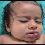
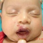
Haemangiomas on the lips
Haemangiomas on the lips often become ulcerated, and because ulcers are so painful, the child may not want to feed. Giving your child pain relief, before feeding can help. It can also help to put some Vaseline on the teat of the bottle (or around your nipple if you are breast feeding) will reduce friction and make feeding less painful.
Treatment options
There are lots of treatment options available; which option is suggested depends on the size and location of your child’s haemangioma.
Haemangiomas are painful when they are ulcerated, so your child may need regular pain relief.
Antibiotics
Antibiotics are recommended when an infection of an ulcerated haemangioma is suspected. These are usually given in the form of a cream or ointment to put directly on the ulcerated area, but widespread infection may need antibiotics in the form of tablets or a liquid as well. Occasionally, a child might need intravenous (into a vein) antibiotics and a stay in hospital.
Steroids
Steroids may be needed when the haemangioma is near the eye, lips, or nappy area to limit further growth. They are usually given as tablets or a liquid, and need to be given exactly according to instructions. When the haemangioma has shrunk enough, the dosage is gradually reduced, as it can be dangerous to stop steroids suddenly.
Laser Treatment
Laser treatment is sometimes used in the treatment of haemangiomas. It can be useful if used early in the development of flat haemangiomas, as it reduces the redness and also reduces the risk of ulceration. Laser treatment is also helpful if an ulcer has already developed, as it reduces the pain of the ulcer, makes it heal faster and reduces the risk of infection.
Immunosuppressive Medicines
A medicine called vincristine is occasionally used to treat specific types of haemangioma in infancy. This medicine is usually used to treat certain forms of cancer and leukaemia, but the doses used to treat these specific haemangiomas are much smaller. It is usually given alongside steroid medicines, particularly if they have not reduced the swelling.
Embolisation
Embolisation is a procedure to ‘block off’ certain blood vessels by injecting them with a ‘glue-like’ substance or beads that stop the blood flowing through them. Generally, this is only used when haemangiomas are very troublesome and other treatments have had little effect.
Surgery
Sometimes, surgery is an option for removing haemangiomas. The surgeon removes the haemangioma tissue and joins the healthy skin together. There will always be a scar with this sort of operation, but it varies depending on the location and size of the haemangioma. Sometimes, it is better to wait until the haemangioma has resolved completely, and see whether surgery is needed at that point.
What are the long term effects of a haemangioma?
Most haemangiomas will have disappeared completely by the age of five to seven years. Large haemangiomas may continue to get smaller until your child is about eight to ten years old.
Depending on the size and location of the haemangioma, there may be little sign it ever existed. Occasionally the affected area of skin might be a bit lighter in color than the rest of your child’s skin.
Large haemangiomas may distort the surrounding skin and even when they disappear, leave behind an area of stretched skin that looks puckered or wrinkled. This can often be improved with cosmetic surgery. Some children may have an uneven skin texture once the haemangioma has resolved. Some children may have some leftover fatty tissue.
GUJARATI
હિમેન્જયોમા (લોહીની નળીઓની ગાંઠ)
બાળકનું શરીર બે કોષમાંથી આકાર લેતું હોય છે. આ કોષમાંથી નવા-નવા કોષો ઉદ્ભવીને શરીરના અવયવો બનાવતા હોય છે. એમા લોહીની નળીઓ બનાવતા કોષ પણ હોય છે. અમુક પરિસ્થતીમાં લોહીની નળીઓ બનાવતા કોષ છુટા પડી જઈ પોતાનું એક અલગ ઝુમખું અથવા તો ગાંઠ બનાવતા હોય છે. જેને લોહીની નળીઓની ગાંઠ કે હિમેન્જયોમા કહેવામાં આવે છે. હિમેન્જયોમા બાળક જન્મતાની સાથે તુરત જ જાવામાં આવે છે. સામાન્ય રીતે હિમેન્જયોમા ‘લાખુ’ સમજવામાં આવે છે જે ખોટું છે. હિમેન્જયોમા કે લોહીની નળીઓની ગાંઠ પહેલા એક થી દોઢ વર્ષમાં ધીમે-ધીમે વધતી હોય છે અને બે વર્ષની ઉંમર પછી ધીરે-ધીરે તેની જાતે જ ઓછી થવા લાગતી હોય છે. પરંતુ જ્યારે હિમેન્જયોમા શરીરના અગત્યના અવયવો જેમ કે આંખ, નાક, મોઢું, પેશાબનો રસ્તો અથવા કાન વગેરે જગ્યાએ હોય અથવાતો હિમેન્જયોમા જા ઇન્ફેકશન જાણવામાં આવતું હોય તો તેની સારવાર વહેલી કરવાની સલાહ આપવામાં આવે છે. ઘણીવાર હિમેન્જયોમા શરીરના અગત્યના ભાગો ઉપર ખૂબ જ વધારે પડતાં પ્રસરીત હોય છે ત્યારે હિમેન્જયોમા ઓછા કરવા માટે અમુક દવાઓ પણ આપવામાં આવે છે. હિમેન્જયોમા દરેક વખતે સર્જરીથી જ મટતો હોય તેવું જરૂરી નથી. હિમેન્જયોમા અંદર રહેલી લોહીની નળીઓને સંકોચાવા માટે ઇન્જેક્શનનો પણ ઉપયોગ કરવામાં આવે છે. •


HINDI
हिमेंजीयोमा (खून की नलिकाओं की गांठ)
बच्चे का शरीर दो कोशिकाओं से आकार लेता है। इन कोशिकाओं से नयी-नयी कोशिकाएँ बनती हैं, जिनसे शरीर के विभिन्न अंग निर्मित होते हैं। इनमें खून की नलिकाएं बनानेवाली कोशिकाएँ भी होती हैं। कुछ परिस्थितियों में खून की नलिकाएं बनानेवाली कोशिकाएं अलग हो जाती है और एक अलग गुच्छा अथवा गांठ बनाती है। इन्हें खून की नलिकाओं की गांठ अथवा हिमेंजीयोमा कहते हैं। हिमेंजीयोमा बच्चे के जन्म के तुरंत बाद देखा जा सकता है। सामान्यतः हिमेंजीयोमा को ‘जन्म का निशान’ के रूप में समझा जाता है लेकिन यह गलत है। सामान्यतः हिमेंजीयोमा अथवा खून की नलिकाओं की गांठ प्रथम एक से डेढ़ वर्ष में धीरे-धीरे बढ़ती जाती है। और तत्पश्चात, दो वर्ष की आयु के बाद धीरे-धीरे अपने आप ही कम होने लगती है। लेकिन जब हिमेंजीयोमा शरीर के महत्वपूर्ण अंग जैसेकि आँखें, नाक, मुँह, मूत्रमार्ग अथवा कान आदि में होता है अथवा हिमेंजीयोमा में यदि संक्रमण पाया जाता है तो उसका उपचार शीघ्र ही करने की सलाह दी जाती है। कई बार हिमेंजीयोमा शरीर के महत्वपूर्ण अंगों पर बहुत अधिक फैलता है, तब हिमेंजीयोमा को कम करने के लिए कुछ दवाइयाँ भी दी जाती है। यह आवश्यक नहीं है कि हिमेंजीयोमा सर्जरी से हमेशा ठीक होगा। कई बार हिमेंजीयोमा में अंदरूनी खून की नलिकाओं को संकुचित करने के लिए इंजेक्शन का प्रयोग भी किया जाता है। •