Choledochal Cysts in Children
A choledochal cyst is a rare congenital swelling of the hepatic or bile duct of the child’s liver.
- This is the tract that transports bile produced by the cells to the gallbladder and duodenum (the first part of the small intestine).
- These cysts can be intrahepatic, meaning that they occur in the part of the duct located inside of the liver.
- They can also be extrahepatic, meaning part of the bile duct that is located outside the liver.
What does the liver do?
The liver is located in the upper right-hand portion of the child’s abdominal cavity, beneath the diaphragm and on top of the stomach, right kidney and intestines. The liver consists of two main lobes, both of which are made up of thousands of lobules.These lobules are connected to small ducts that connect with larger ducts to ultimately form the hepatic duct. The hepatic duct transports bile produced by the liver cells to the gallbladder and duodenum, which helps to break down fats, preparing them for further digestion and absorption.
All of the blood leaving the stomach and intestines passes through the liver. The liver processes this blood and breaks down the nutrients and drugs in the blood into forms that are easier to use for the rest of the body.
What are the most common varieties of these cysts?
There are five basic types and they’re classified by where they appear.
- Type 1 — A cystic dilation of the extrahepatic bililary duct. This is the most common type, making up about half of all choledochal cysts.
- Type 2 — An abnormal pouch or sac opening from the duct.
- Type 3 —A cyst located within the duodenal wall.
- Type 4 — Swellings of both the intrahepatic and extrahepatic biliary tracts.
- Type 5 —Multiple intrahepatic cysts. This type of clustering of cysts is also known as Caroli’s disease and is the least common type.
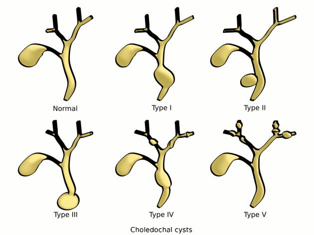 Types of Choledochal Cysts
How common are choledochal cysts?
- Choledochal cysts occur in between 1 and 100,000 and 1 in 150,000 people in Western countries but is much more common in Japan.
- They are three to eight times more common in females than males.
What causes choledochal cysts?
The cause of these cysts is unknown, but researchers believe that their formation is due to an abnormal connection between the pancreatic and hepatic ducts.
This abnormal connection leads to a reflux of pancreatic juice into the biliary tract, which could be responsible for cyst formation.
What are the symptoms of choledochal cysts?
The following symptoms are usually experienced by an older child whose earlier diagnosis of this congenital anomaly may have been missed.
- Abdominal pain in the right upper quadrant
- Jaundice
- Abdominal Mass
- Nausea
- Fever
- Pancreatitis
How are choledochal cysts diagnosed?
Choledochal cysts can be diagnosed prenatally (before birth) on an ultrasound.
After your baby is born, you or your child’s doctor may notice a right upper quadrant mass, with or without jaundice.
Your child will likely undergo a combination of the following tests to verify the diagnosis:
Computerized tomography scan (CT or CAT scan):
A CT scan shows detailed images of any part of your child’s biliary system.
Magnetic Resonance Cholangiopancreatography (MRCP):
Magnetic resonance cholangiopancreatography (MRCP) is a special type of magnetic resonance imaging (MRI) exam that produces detailed images of the hepatobiliary and pancreatic systems, including the liver, gallbladder, bile ducts, pancreas and pancreatic duct.
What is the treatment for choledochal cysts?
Surgery is the only feasible treatment for choledochal cyst. This involves removal of the choledochal cyst, followed by duct reconstruction using a piece of intestine. A number of possible approaches exist, depending on cyst location etc… Sometimes, the appendix is also used as a conduit for reconstruction of the biliary tree.


Without surgery, there is an ongoing risk of other problems:
- Biliary obstruction and stone formation
- Infection of the ducts
- Jaundice
- Cirrhosis
- Perforation of the cyst
Another long-term concern is for malignant degeneration. Choledochal cysts are inflammatory in nature, which makes them at risk for becoming cancerous if left untreated.
Outcome for children with choledochal cysts after surgery
Children can lead a normal life after surgery for choledochal cysts. There are no issues related to digestion or their growing up. We normally follow then up after surgery with ultrasound scan after 6 months. If required blood tests are also carried out on follow up.